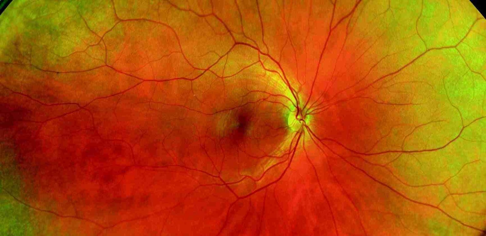
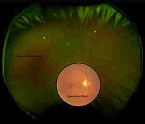
As optometrists, we are so privileged to be supported by the latest eyecare technology, including the Optos Ultra-Widefield camera. This tool allows us to take a single retinal photo that can image up to 200 degrees of the retina (compared to standard 45 degrees). It is as simple as taking a photo on your phone and doesn’t require eyedrops, which can blur your vision. Ocular health issues can often be identified without any presenting complaints or warning signs to you, our patients.
Below outlines an interaction with a recent patient who was interested in updating glasses following a check 18 months ago.
Patient Profile
- 60 year old Male – no significant history.
- Upon questioning, mentioned that several months prior he had noticed some floaters which have since disappeared.
The Challenge
Given the long-standing nature of the symptoms and considering it was not a current concern to the patient, Nick had the option of:
- Measuring the spectacle prescription, dispensing new glasses and then dilating the patient’s eyes which would have made for a much longer appointment and meant the patient was left with blurry vision and would be unable to drive or work comfortably for a few hours following.
- Measuring the spectacle prescription, dispensing new glasses and recommending the patient return for a dilated fundus examination which are often overlooked by patients in their busy lives.
- Forgetting the patient’s wishes of new spectacles, and dilating the pupil, to focus on the floaters which is a larger concern for optometrists.
- Measuring the spectacle prescription, dispensing new glasses and recommending an ultra-widefield image of the eye as part of the routine examination to make sure the floaters weren’t associated with damage to the back of the eye, with the option to recommend the dilating drops depending on the results.
Nick chose option 4 – Implementation of Optos Widefield Imaging
- Comprehensive Retinal View: Unlike traditional imaging methods that might capture only a portion of the retina, Optos Ultra-Widefield Imaging provided a detailed, larger view of the patient’s retina. This identified two retinal tears in the periphery.
- Patient Education: The high-resolution images provided a clear and detailed visual representation of the concern for the patient, allowing understanding and good compliance to treatment.
- Enhanced Monitoring: The widefield images serve as good baselines for subtle comparisons over time, just like conventional retinal photography.
- Non-Invasive and Efficient: Optos Ultra-Widefield Imaging is non-invasive and relatively quick, making it more comfortable for patients compared to some traditional examination techniques. This efficiency also allows for more comprehensive examinations within the same appointment time.
Outcomes
Following the introduction of Optos Widefield Imaging:
- Improved Diagnosis: Nick was able to identify two retinal tears without the need for blurring, dilating drops.
- Better Management: The patient understood the concern and was able to be referred for vision saving laser retinal treatment the following day.
- Enhanced Patient Satisfaction: A routine examination detected and prevented further visual loss for the patient whilst also being able to update to some lovely frames.
The use of Optos Ultra-Widefield Imaging is not limited to patients with acute concerns – it applies to everyone who wants a comprehensive check of their eyes. It can detect general health issues including identifying different cardiovascular problems as part of routine checks.
Book your appointment today and discuss how Optos imaging may be of benefit in your examination.